- GE HealthCare recently announced the submission of 510(k) to the U.S. FDA, seeking clearance for the Photonova Spectra, its new photon-counting computed tomography system, marking a significant step in GE HealthCare’s ongoing CT research and development efforts.
- The system aims to enable ultra-high-definition chest scans in as little as one second, supporting clinicians in evaluating fine details with exceptional clarity.
- Photon-counting CT directly counts individual X-ray photons and records their energies, enabling multi-energy information from a single scan.
- In cardiology, Photonova Spectra is designed to achieve robust and dose-conscious ultra-high-definition heart imaging with wide volumetric coverage.

GE HealthCare recently announced the submission of 510(k)s to the U.S. FDA seeking clearance for the Photonova™ Spectra1, its new photon-counting computed tomography (PCCT) system, marking a significant step in GE HealthCare’s ongoing CT research and development efforts.
Photon-counting CT directly counts individual X-ray photons and records their energies, enabling multi-energy information from a single scan. This approach helps provide excellent contrast-to-noise ratio at comparable dose, and the potential to sharpen fine structural details, and generate quantitative material maps that can empower clinicians to characterize disease with great confidence. GE HealthCare’s Deep Silicon design, Photonova Spectra (510(k) pending at the U.S. FDA; Not available for sale) builds on this foundation with an edge-on silicon detector architecture that is designed with the goals of high spectral precision even at high count rates, combined with small, highly efficient pixels.
In cardiology, Photonova Spectra is designed to achieve robust and dose-conscious ultra-high-definition heart imaging with wide volumetric coverage. The system aims to enable ultra-high-definition chest scans in as little as one second, supporting clinicians in evaluating fine details with exceptional clarity. The technology is designed to help clinicians directly visualize small fractures and bone marrow edema, supporting detailed clinical assessments for orthopedic care.
In this article, we will examine how CT technology works, explore the fundamentals of photon-counting detection, and reveal why we believe GE HealthCare’s Deep Silicon approach represents a new generation of spectral photon counting CT, powered by 8-bins of energy resolution.
The foundations of traditional CT
Computed tomography (CT) works by generating X-rays in a high-voltage tube, sending them through the patient, and recording the attenuated signal with a detector. A tungsten target struck by an electron beam produces X-rays of specific tube potentials, typically between 80 and 140 kilovolts peak (kVp). Each emitted photon, however, carries its own energy value, measured in kilo-electronvolts (keV), which can be up to the tube potential. As the gantry spins, X-rays pass through the body and are absorbed by tissues depending on their thickness and density, following the Beer–Lambert principle, which describes how intensity decreases as radiation travels through matter. This means denser tissues such as bone absorb more X-rays and appear whiter on the image, while softer tissues or air-filled lungs absorb less and appear darker.
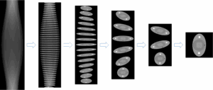
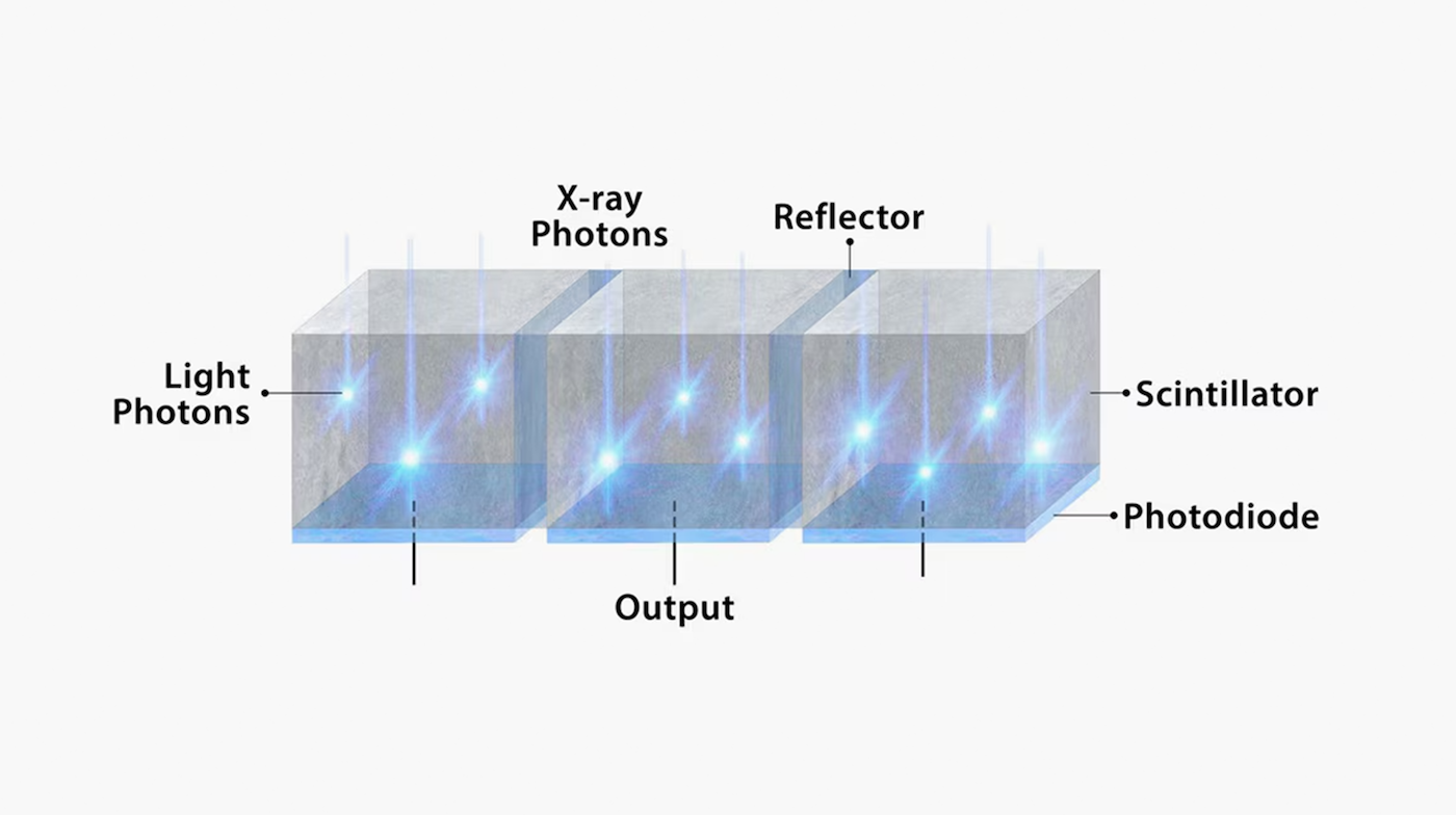
The detector measures the transmitted intensity and converts it into electronic signals, which are reconstructed into detailed cross-sectional images. Conventional scintillator-based CT detectors first convert X-rays into light and then light into electrical signals. They use reflective foils between roughly millimeter-sized pixels to keep the light generated by X-ray absorption from spreading sideways. These foils are essential for accurate light detection but occupy space that does not contribute to capturing X-rays.
The reconstruction process is a formidable computational challenge: tens of thousands of samples from each of half a million pixels yield billions of equations that the system must solve to form a single three-dimensional image volume. Algorithms such as filtered back projection use Fourier mathematics to reconstruct these signals into a faithful depiction of anatomical structures. In essence, CT turns invisible radiation measurements into visual representations of organs and tissues with astonishing speed and precision.
The clarity and diagnostic value of a CT image depend on spectral, spatial, and temporal resolution. Spectral resolution refers to how well the system distinguishes materials by their X-ray absorption at different energies, information essential for increased conspicuity of iodinated contrast and for characterizing pathological tissues. Spatial resolution defines how sharply fine details appear and is governed by the focal spot, the detector pixel size, and the modeling power of the reconstruction algorithm. Temporal resolution determines how effectively the scanner freezes motion, especially in the heart or lungs, where rapid rotation and motion-correction algorithms aim to prevent blurring. Photon counting does not itself change temporal resolution; fast rotation and motion correction are separate system attributes, not inherent to but inherited by PCCT.
The emergence of photon counting
Photon-counting detectors (PCDs) are designed to detect and count individual X-ray photons directly. When a photon enters a semiconductor sensor such as cadmium telluride (CdTe), cadmium zinc telluride (CZT), or silicon, it generates a small electric charge pulse proportional to its energy. Ultra-fast application-specific integrated circuits (ASICs) amplify and shape these pulses, while on-pixel comparators compare each event to specific energy thresholds, creating multiple data streams called energy bins that record photons by energy level. This direct conversion aims to preserve each photon’s energy information and reduce the impact of electronic noise, allowing quantitative multi-energy measurements from a single scan.

What’s innovative with Photonova Spectra (510(k) Pending at the U.S. FDA; Not available for sale)
Photonova Spectra’s design uses silicon as a detector material. Silicon was chosen as the foundation for GE HealthCare’s Deep Silicon detector technology because of its high intrinsic energy resolution, as well as its exceptional purity, stability, and scalability. The uniform crystal structure and low defect density of silicon supports the design intention of consistent detector performance across large areas.
Silicon has a lower atomic number and density than CdTe or CZT, meaning it absorbs X-rays less efficiently in traditional face-on configurations. GE HealthCare’s edge-on geometry is designed to address this by dramatically increasing the X-ray path length through the material. This geometry boosts absorption efficiency while keeping the material thin and manageable for production. The result is a detector that is designed to combine exceptional spectral precision and high count-rate performance.
Photonova Spectra introduces four foundational innovations that aim to support precise and high-quality images: multiple energy bins for robust material characterization, ultra-small effective pixel size through minimization of charge sharing, low-noise direct conversion to support dose efficiency, and avoiding pile-up through multiple depth segments.
- Eight energy bins for robust material characterization
Photon counting is designed to measure every individual X-ray photon and records its energy across several discrete ranges known as energy bins. These multi-threshold measurements are then decomposed into a small set of basis materials commonly water and iodine before image reconstruction. The resulting data is transformed into virtual monoenergetic images and quantitative maps of iodine or other materials. With eight distinct clinical energy bins, GE HealthCare’s Deep Silicon detector technology is designed to leverage the intrinsic sharpness of the silicon photopeak, which exhibits minimal overlap among adjacent energies. This high precision spectral separation aims to enhance lesion characterization, provide impressive iodine contrast, and support precise quantitative imaging while maintaining a low radiation dose. It also aims to create enhanced contrast between soft tissues, such as between white and gray matter in the brain. Because iodine absorbs more strongly at lower photon energies, the scanner is designed to emphasize low-energy bands to highlight iodine-enhanced tissues, with the goal yielding conspicuity of subtle lesions and aiming to help improve low-contrast detectability. - Ultra-high spatial resolution through minimization of charge sharing
A long-standing technical hurdle in photon-counting CT has been charge sharing the unwanted spreading of an X-ray’s electric charge across multiple pixels, blurring spatial detail and increasing the effective pixel size beyond its nominal size. GE HealthCare’s edge-on silicon architecture is designed to address this challenge through minimizing the amount of charge sharing. X-rays enter along the wafer edge to extend the photon path, and the thin wafer design controls the charge spread. In addition, thin tungsten foils between silicon sensors block stray scatter and limit crosstalk. These innovations aim to maintain both spectral precision and sharp spatial resolution, preserving the benefit of the eight energy bins and the small pixel size. - Low-noise direct conversion: protecting low-dose performance
Traditional CT detectors may experience electronic noise and random background signals that impact image quality, especially at low radiation doses. Photon-counting CT has the goal of addressing this limitation by directly converting X-rays into electrical signals and counting individual photons, without the intermediate light conversion step. Operating above a low-energy threshold, the system is designed to remove nearly all electronic noise, leaving only true photon statistics. This means the scanner is designed to generate images with good contrast-to-noise ratio even at low dose levels. - Pulse pile-up management for maintaining accuracy at high photon flux
A key technical challenge in photon-counting CT is pulse pile-up, which happens when multiple photons arrive at a detector pixel within billionths of a second, causing their electrical signals to overlap. This overlap can produce false readings or energy distortion at high scan speeds. Photonova Spectra’s Deep Silicon detector is designed with this in mind, with its edge-on, multilayer design, where each pixel is divided into multiple depth segments that function as independent detection zones. This structure distributes the photon flux across layers, aiming to reduce the likelihood of overlapping events and helping support accurate photon energy measurement even under high clinical X-ray flux.
Together, these four innovations define the advancements with Photonova Spectra, which aims to provide accurate material characterization, consistently sharp, detailed imaging across the body, and exceptional low-dose and high-flux performance.
By combining a distinctive detector design, the excellent material properties of silicon, and advanced algorithms, GE HealthCare’s Deep Silicon scanner is designed to achieve high count rates, accurate spectral discrimination, and ultra-fine spatial resolution in a single system.