The GE HealthCare Technology and Innovation Center (HTIC) serves as a centralized research and development hub for the company. This article examines how HTIC’s MRI and Superconducting Magnet Research team—comprising approximately 20 specialists across physics, chemistry, computational sciences and various branches of engineering—has systematically addressed fundamental limitations in brain imaging through a family of high-performance MRI systems. More specifically, we trace the evolution of their technologies across successive investigational devices—from the investigational Compact 3 Tesla (T) system‡ developed in 20151, through an investigational gradient insert‡ developed in 20192, and the investigational Compact 7T scanner‡ in 20253.
The four essential subsystems of an MRI scanner
To appreciate these innovations, it is essential to understand how MRI creates images. At a broad level, an MR system has four components:
- The Superconducting Magnet is a bedrock of MRI. This incredibly powerful magnet—about 60,000 times stronger (for 3T) than Earth’s magnetic field—aligns hydrogen nuclei in your body’s water molecules like compass needles. What makes this practical is superconductivity: special wires (niobium-titanium) cooled to extremely low temperatures – 4.2 Kelvin (that is, -452 °F!) – using liquid helium. At this temperature, the wires lose virtually all electrical resistance. Once a current is injected into these near-zero-resistance coils, it can be sustained without additional power, creating a strong, persistent magnetic field essential for MRI.
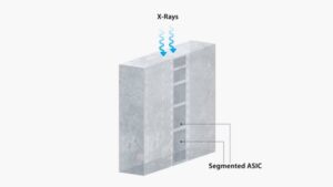
- The RF Transmit System disrupts this alignment of hydrogen nuclei to create detectable signals. It sends radio frequency (RF) waves that tip the aligned hydrogen nuclei (i.e., spins) in your body’s water molecules, which sets them up for subsequent manipulation to achieve different types of image contrasts. However, these radio waves also generate electric fields that can heat tissues over time—this is measured by the Specific Absorption Rate (SAR), which tracks how much energy your body absorbs. At higher magnetic field strengths (e.g., 7T, which is about 140,000 times stronger than Earth’s magnetic field), more advanced multi-channel RF transmit architectures and software are needed to minimize any local, concentrated heating in tissues.
- The Gradient System provides spatial encoding of spins—essentially MRI’s GPS. By creating controlled magnetic field variations across the imaging volume, each location is encoded with a unique frequency and phase signature. Imaging performance is enhanced with higher gradient amplitudes (strength of gradient fields) and slew rates (switching speed of gradient fields).
- The RF Receive System captures the faint MR signals emitted as the spins precess – the wobbling motion of hydrogen nuclei spins when they are placed in a strong magnetic field. Arrays of sensitive antennas, called receive coils, surrounding the anatomy collect these signals. Advanced software then processes the raw data to reconstruct detailed medical images.
Establishing foundational technologies for MRI
HTIC’s MRI laboratory has shaped the field since the 1980s, transforming early 0.5-0.7T scanners with limited image quality into today’s sophisticated systems. The lab pioneered whole-body MRI at higher magnetic fields, establishing 1.5T as the enduring clinical standard worldwide.
The laboratory developed groundbreaking technologies that transformed MRI’s clinical capabilities. The RF birdcage coil4 works like an antenna that sends and receives radio waves uniformly throughout the body, ensuring consistent image quality across the entire scan. Phased array RF receive coils5 function like multiple antennas working together, capturing clearer signals that produce sharper images in less time. Field shaping arrays6 correct the radio wave patterns that can become distorted in powerful magnets, while dynamic shimming7 actively adjusts the magnetic field during scanning to prevent image warping and ensure anatomical accuracy.
HTIC has pioneered another revolution: dramatically reducing MRI’s dependence on liquid helium. Traditional systems require 1500-2000 liters of liquid helium, which is a non-renewable resource that is increasingly scarce and expensive. HTIC’s 2011-2012 breakthrough in advanced cryogenics systems technology reduced liquid helium requirements.
Working backwards from clinical outcomes: How HTIC solves complex problems
HTIC engages clinical collaborators to advance the frontiers of medical technology to deliver transformative outcomes for patients. For example, through close collaborations, a critical challenge was identified: imaging brain microstructure to detect disease earlier, identify subtle injuries, and monitor treatment response with unprecedented precision.
To address this, HTIC is exploring advanced imaging techniques such as:
- Oscillating Gradient Spin Echo (OGSE): which enhances sensitivity to microstructural changes, which has the potential to offer insights critical for tumor grading and treatment planning.
- Axonal Diameter Imaging (AXDI): that enables evaluation of white matter integrity, which may provide valuable information on brain development, learning, and changes in connectivity due to disease or rehabilitation.
- Simultaneous Coherent and Incoherent Motion Imaging (SCIMI): which tracks neurofluid dynamics involved in waste clearance and inflammation—processes which may be linked to conditions like hydrocephalus, Alzheimer’s disease, and traumatic brain injury.
These techniques use special types of MRI scans that produce information about tiny structures less than 10 micrometers (about 1/10 the width of a human hair) and detect very slow fluid movements of less than 100 micrometers per second. However, capturing this level of detail requires extremely powerful gradients (≥200 mT/m strength, >500 T/m/s speed) that full-body MRI scanners cannot achieve as they would cause uncomfortable peripheral nerve stimulation in the body. Clearly, a new paradigm was needed.
The breakthrough insight: Less is more
The team recognized that in a smaller, head-only system, stronger and faster switching gradient fields can be used because of the higher peripheral nerve stimulation threshold in the head. The team also recognized early on that technology excellence alone isn’t enough. To truly transform clinical practice, a head-only scanner must not only surpass the performance limits of traditional MRI machines – a head-only scanner must also be easy to use and cost-effective to install and operate. This realization shaped HTIC’s three-dimensional approach to innovation in the development of investigational neuroimaging systems—balancing usability, performance, and scalability.
Building a framework: Three Dimensions of Innovation
The team’s strategic framework for neuroimaging innovation operates along three essential dimensions that must work in harmony for clinical success.
- The first dimension, access, recognizes that even the most advanced technologies will have limited impact if hospitals cannot install or afford it, e.g., traditional 7T scanners demand purpose-built facilities with reinforced floors for 20-40 tonne magnets and massive cooling infrastructures.
- The second dimension, gradient performance, addresses the fundamental limitation preventing visualization of brain microstructure: the PNS thresholds for whole-body gradients significantly limits the usable range of gradient performance compared to head-only gradients.
- The third dimension addresses the engineering challenges in increasing the static magnetic field in low cryogen magnets to produce images with higher signal-to-noise ratio (which leads to higher quality images).
The team places an equally important, overarching emphasis on the development of intelligent agents and advanced applications that can leverage the increased gradient performance and field strengths to potentially enhance the quality of information for diagnosis and therapy planning.
From vision to reality
The evolution of HTIC’s neuroimaging systems demonstrates how systematic innovation progressively addresses these dimensions. The investigational Compact 3T system (2015) proved that low-cryogen technology could work reliably in a clinical setting. The development of this system was funded by NIH, which chartered the team to create a high-performance 3T neuroimaging scanner that was easier and less costly to install and maintain. Weighing 2 tonnes – less than one-third of conventional whole-body 3T scanners – it uses an innovative magnet with less than 12 liters of sealed liquid helium versus 1500-2000 liters in conventional systems. The gradient coil can produce a maximum gradient strength of 80 mT/m and a gradient field switching rate of more than 700 T/m/s, resulting in higher quality images with significantly less geometric distortions.
An investigational gradient insert (2019), funded by the Congressionally Directed Medical Research Program (CDMRP), represented the next evolutionary step, addressing access and gradient performance simultaneously. The design has remarkable power efficiency that is significantly higher than conventional whole-body systems, enabling 300 mT/m strength and 750 T/m/s using a 2 MVA gradient driver per axis. This enables fast ultra-detailed brain scans while revealing cellular-level information that is invisible to standard clinical MRI scanners. The additional information may help clinicians better understand brain function, structure and pathologies. Notably, this capability could be attained without a need to upgrade existing power infrastructure that already supports 3T whole-body scanners.
The investigational Compact 7T system (2025) culminates HTIC’s strategic vision, addressing all three dimensions. Drawing on invaluable learnings gleaned from the development of the Compact 3T system in 2015, the team developed an investigational 7T head-only system that can fit within a standard 3T whole-body magnet room, reducing the need for costly infrastructure upgrades. Developed through NIH funding with Mayo Clinic and UCSF, the system weighs just 8 tonnes – compared to the 20-40 tonnes typical in whole-body 7T systems – and uses 18 liters of permanently sealed helium, a dramatic reduction from the 2000 liters required in conventional 7T systems. Its advanced cryogenics systems eliminate complex venting systems that can increase installation costs significantly, especially in complex building structures. The high-performance gradient coils, combined with 7T’s excellent signal quality, could potentially enable research-grade brain imaging in clinical practice one day.
HTIC’s approach offers a powerful blueprint for advancing medical technology—beginning with the identification of critical clinical needs, addressing foundational scientific challenges, and ensuring that innovations are practical, scalable, and could be ready for real-world adoption. By balancing near-term clinical needs with transformative long-term research, the team continues pushing boundaries while staying grounded in translating innovations to improve patient care.
‡ Prototypes in development that represent ongoing research and development efforts. These technologies are not products and may never become products. Not for sale. Not CE marked. Not cleared or approved by the US FDA or any other global regulator for commercial availability.
- Foo TKF, Laskaris E, Vermilyea M, Xu M, Thompson P, Conte G, Van Epps C, Immer C, Lee SK, Tan ET, Graziani D, Mathieu JB, Hardy CJ, Schenck JF, Fiveland E, Stautner W, Ricci J, Piel J, Park K, Hua Y, Bai Y, Kagan A, Stanley D, Weavers PT, Gray E, Shu Y, Frick MA, Campeau NG, Trzasko J, Huston J 3rd, Bernstein MA. Lightweight, compact, and high-performance 3T MR system for imaging the brain and extremities. Magn Reson Med. 2018 Nov;80(5):2232-2245. ↩︎
- Foo TKF, Tan ET, Vermilyea ME, Hua Y, Fiveland EW, Piel JE, Park K, Ricci J, Thompson PS, Graziani D, Conte G, Kagan A, Bai Y, Vasil C, Tarasek M, Yeo DTB, Snell F, Lee D, Dean A, DeMarco JK, Shih RY, Hood MN, Chae H, Ho VB. Highly efficient head-only magnetic field insert gradient coil for achieving simultaneous high gradient amplitude and slew rate at 3.0T for brain microstructure imaging. Magn Reson Med. 2020 Jun;83(6):2356-2369. ↩︎
- Wu A, Ricci J, Conte G, Van Epps C, Xu M, Bai Y. Design and construction of a low-cryogen, lightweight, head-only 7T MRI magnet. IEEE Trans on Appl Supercond. 2024 Aug;34(5):1-5. ↩︎
- Hayes CE, Edelstein WA, Schenck JF, Mueller OM, Eash M. An efficient, highly homogeneous radiofrequency coil for whole-body NMR imaging at 1.5 T. Jour Mag Reason. 1985; 63: 622–628. ↩︎
- Roemer PB, Edelstein WA, Hayes CE, Souza SP, Mueller OM. The NMR phased array. Magn Reson Med. 1990 Nov;16(2):192-225. ↩︎
- Hancu I, Lee SK, Dixon WT, Sacolick L, Becerra R, Zhang Z, McKinnon G, Alagappan V. Field shaping arrays: a means to address shading in high field breast MRI. J Magn Reson Imag. 2012 Oct;36(4):865-72. ↩︎
- Lee SK, Tan ET, Govenkar A, Hancu I. Dynamic slice‐dependent shim and center frequency update in 3 T breast diffusion weighted imaging. Magn Reson Med. 2014 May;71(5):1813-8. ↩︎