

Traditional management of heart disease remains largely reactive. Cardiovascular disease is the leading cause of death globally, taking approximately 17.9 million lives each year. At the same time, heart failure is often identified only after patients have already reached an acute care setting. Recent research suggests that 38 percent of newly diagnosed heart failure patients were diagnosed in acute care settings, and 46 percent of those patients had potential heart failure symptoms in the prior six months.1
Many patients show signs of cardiovascular disease before diagnosis, but the tools needed to evaluate cardiac function are not always available where they first seek care. Symptoms such as shortness of breath, fatigue, edema, chest discomfort, hypotension, and reduced exercise intolerance may present in primary care clinics, urgent care centers, emergency departments, ambulances, rural hospitals, or community settings. And yet, conventional echocardiography often requires specialized equipment, trained sonographers, expert interpretation, and dedicated scheduling pathways.
As a result, delays can arise between clinical suspicion and diagnostic confirmation.
To give just one example, consider left ventricular ejection fraction (LVEF), one of the most commonly used measurements in cardiovascular medicine. LVEF quantifies how effectively the heart pumps blood and serves as a reliable indicator to inform treatment planning across a wide range of cardiac conditions.
Current echocardiography guidelines emphasize accurate quantification of LVEF because numerous therapeutic decisions rely on specific ejection fraction (EF) thresholds. Whether initiating medical therapy, evaluating heart failure severity, determining eligibility for advanced interventions, or monitoring disease progression, clinicians frequently depend on an accurate assessment of ventricular function.
Echocardiography can provide these answers, but access often depends on where the patient is being evaluated and how quickly imaging resources are available. As a result, clinically important information about cardiac function may not be available when critical decisions are being made.
Bringing ultrasound to the point of care with Vscan Air SL
Vscan Air SL was designed to address this challenge by bringing cardiac imaging earlier in the patient care path.
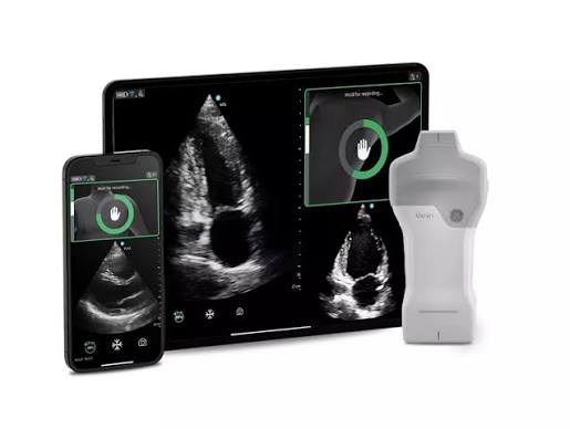
The system combines a wireless handheld ultrasound probe with smartphone or tablet connectivity, enabling clinicians to perform cardiac and vascular assessments at the point of care. Rather than requiring access to a dedicated imaging suite, clinicians can acquire ultrasound images wherever patients are being evaluated.
The device incorporates a dual-probe design. One side contains a sector phased-array transducer, a type of ultrasound probe that emits and steers sound waves electronically to create a wedge-shaped image. This design is particularly well suited for cardiac imaging because it can image deep structures through the narrow acoustic windows between the ribs.
The other side contains a linear-array transducer, which produces a rectangular image and is optimized for superficial structures located closer to the skin, such as blood vessels in the neck, arms, and legs. This allows clinicians to transition between cardiac and vascular examinations without changing equipment.
Proprietary imaging technology delivers high-intensity signal processing for exceptional penetration, resolution and sensitivity in imaging performance. Cloud connectivity, remote image review, live collaboration tools, and bedside documentation further extend the platform’s capabilities, enabling imaging to be acquired, reviewed, shared, and documented within the clinical workflow.
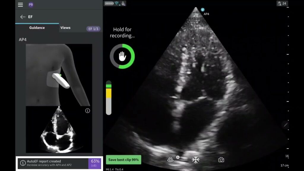
However, one of the most remarkable aspects of this system is the software that offers even more to users. Acquiring an image is only the first step; the greater challenge is getting a reliable image to support a reliable assessment of cardiac function by a provider at the point of care. Caption AITM can help address this challenge through deep learning algorithms designed to guide image acquisition and estimate cardiac performance in a way that closely mirrors how experienced echocardiographers visually assess the heart.
Deep learning that mimics the human eye
LVEF describes how effectively the left ventricle pumps blood during each cardiac cycle by measuring the proportion of blood ejected during systole relative to the volume present at end-diastole. As the ventricle fills with blood during diastole and contracts during systole, EF provides a quantitative measure of global systolic function; for example, if the ventricle contains 100 milliliters of blood before contraction and 40 milliliters remain afterward, 60 milliliters have been ejected, resulting in an ejection fraction of 60 percent.
Caption InterpretationTM AutoEF was developed to automate and standardize LVEF visual estimates at the point of care. The most significant technical innovation behind AutoEF is how it approaches ejection fraction estimation differently from traditional echocardiographic methods.
Historically, EF measurement has depended on endocardial border detection. The endocardium forms the inner boundary of the ventricular cavity. Conventional software attempts to identify this border at end-diastole and end-systole, reconstruct ventricular geometry, estimate chamber volumes, and calculate ejection fraction from the resulting volume measurements.
While conceptually straightforward, this process presents substantial technical challenges. Ultrasound images frequently contain acoustic shadowing, speckle noise, dropout artifacts, and incomplete visualization of cardiac structures. Patient body habitus, image quality, and pathological remodeling can further complicate border identification. Even among experienced readers, these factors contribute to variability in measurements.
To overcome these challenges, the AI development team proposed a fundamentally different solution. Instead of asking a computer to identify every ventricular border and calculate ventricular volume, could a computer be trained to recognize the degree of ventricular contraction directly?
This approach mirrors how experienced echocardiographers often estimate ventricular function during routine clinical practice: by recognizing patterns of cardiac motion rather than consciously tracing every border. Furthermore, they look to changes in cavity size, wall motion, and overall contractions throughout the cardiac cycle.
The framework provides the conceptual basis for learning the relationship between visual contraction patterns and ejection fraction.
Letting the algorithm work it out
The algorithm was trained using more than 50,000 echocardiographic studies representing a broad range of image quality, patient anatomy, and cardiovascular pathology.
Each study contained standard echocardiographic views alongside ejection fraction measurements generated through conventional clinical workflows. Over repeated training iterations, the neural network learned which features and motion patterns, and temporal characteristics were associated with specific ejection fraction values.
Notably, the developers did not explicitly instruct the algorithm regarding which structures to track or which features to prioritize. No rules were provided that specified how ventricular walls should be followed throughout the cardiac cycle. The neural network was allowed to derive its own internal representation of the visual information most predictive of ventricular function.
The resulting system can estimate ejection fraction using any combination of standard echocardiographic views, including the apical two-chamber (AP2), apical four-chamber (AP4), and parasternal long-axis (PLAX) views, that meet quality requirements. It also provides an expected error range and a confidence estimate, allowing clinicians to understand not only the predicted value but also the reliability of the prediction.
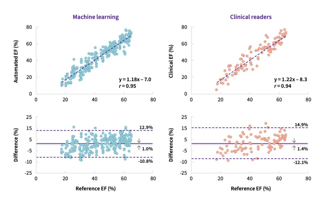
Validation studies demonstrated strong agreement between AutoEF and expert reference measurements. In one study, automated measurements achieved a correlation coefficient of 0.95 with expert-derived reference values, performance comparable to conventional clinical measurements.2
These results suggest that clinically useful assessment of ventricular function can be achieved without requiring explicit ventricular border tracing, opening the possibility of more accessible LVEF estimates across a broader range of care settings.
By combining handheld ultrasound, AI-guided image acquisition, and automated interpretation, technologies such as Vscan Air SL and Caption AI extend aspects of specialized echocardiographic expertise beyond traditional imaging departments, enabling earlier recognition of criteria like ventricular dysfunction.
For clinicians, this provides cardiac assessment support in settings where expert sonographers and cardiologists may not be immediately available. Importantly, these tools are not intended to replace comprehensive echocardiography or specialist interpretation, but rather to bring focused cardiac assessment closer to the patient and the point of clinical decision-making, where earlier intervention can have the greatest impact.
- Journal of the American Heart Association: “Disparity in the Setting of Incident Heart Failure Diagnosis ↩︎
- Internal GE HealthCare Research; Data on file ↩︎