Recently at RSNA 2025, Dr. Thomas Foo became part of a select club: the Academy for Radiology and Biomedical Imaging Research’s Council of Distinguished Investigators, a network of more than 500 imaging scientists.
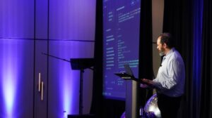
Foo, the chief scientist at GE HealthCare in Niskayuna, N.Y., was named a 2025 Distinguished Investigator, an honor the Academy reserves for researchers whose work advances medical imaging in ways that matter in practice.
Foo’s entry into medical imaging began as an intellectual detour. As an undergraduate at Kenyon College, a small liberal-arts school in Ohio where he studied physics and mathematics, he met a new faculty member trained in medical radiological physics, a path that stood apart from the department’s usual gravitational pull toward high-energy and theoretical physics. The idea that physics could be applied directly to medicine, and that MRI offered a non-ionizing window into soft tissue with extraordinary contrast, hooked him early. He went on to earn a PhD in medical physics at the University of Wisconsin, Madison, choosing it in part because the campus was about to receive one of the first commercial MRI systems in the world, and he wanted to learn the field as it was taking shape.
There, he worked alongside early MRI pioneers, and a brief proposal he wrote became a funded collaboration among U Wisconsin – Madison and GE HealthCare. After graduate school, he joined GE, where fast imaging projects led into cardiac and vascular MRI, followed by a decade of clinical-facing work in the Washington, D.C. area.
Those years, split between engineering and bedside reality, sharpened his focus on what imaging has to deliver to count as progress, not just performance. In this interview, Foo talks about what separates research that changes practice from research that merely improves tools, why MAGNUS required rethinking the hardware itself, and what he believes must happen for advanced imaging to become more widely accessible.

Q: In your view, what distinguishes research that merely advances technology from research that transforms a field?
A: Research can take many forms, but the thing that matters most to me is impact and significance. I was told early on by someone very senior that 90 percent of what you do is project selection. If you select a good project, everything is great. If it’s a bad project, you’re spinning the wheels in the mud.
My background probably shaped this worldview. I’m not one of those people who came up through hardcore engineering from day one. I went to school for liberal arts, and I was first introduced to MR by a faculty member who didn’t fit the traditional physics mold. He had trained in medical radiological physics, and those conversations opened my eyes to the idea that physics could be used directly in medicine.
My liberal arts background helped. We were trained to think, to arrange thoughts, to argue positions, to look at a problem from more than one angle. That turns out to be useful in research because you’re constantly testing your own assumptions.
If you want a practical filter for project selection, I’d ask you to put yourself in the patient’s shoes. Patients want to know what’s wrong, whether they’re getting better or worse, and they want the whole process to be as simple as possible. They want workflows to be reasonably quick, painless, non-interventional, and accessible.
And that’s a great lens to inform your research. Innovations that aim to reach a broad population can have meaningful impact. If something only reaches a small fraction of the population, it may be more limited in its overall mission impact.
Q: The MAGNUS head-only 3T gradient system demonstrated what’s possible when performance meets purpose; looking back, what were the key scientific or strategic decisions that turned it from an ambitious experiment into a platform with translational potential, and what lessons from that journey would you share with researchers working at the edge of what’s technically feasible?
A: One of the big shifts for me happened when I moved from working mainly on applications to working on the platform itself. On a commercial scanner, a lot of the time you’re like someone cooking dinner: you open the fridge, see what’s in there, and make whatever you can with what you’ve got. You can cook a decent meal on a hot plate. But if you’ve got a chef’s kitchen with the right equipment, you can do much more. At the research center, I had the chance to build a better kitchen, not just cook with whatever was already in the fridge.
That mindset was central to how we approached MAGNUS. We looked at the state of research on standard whole-body platforms and saw a lot of incremental work, the same basic results with minor twists. If you want a meaningful advance, we saw that we had to change the hardware.
The other key decision was zeroing in on the right focus area. Whole-body scanners are powerful, but if you try to build something that does everything, you don’t do everything that well. We chose to focus on the brain, because brain injury and neuropsychiatric disease carry significant human and economic cost, and because brain imaging still has room for further improvement.
We pursued a head-only 3T system with high-performance gradients designed to deliver substantially higher gradient performance while maintaining a practical power profile. That was a hard sell at first.
People were comfortable with whole-body systems, and a lot of people remembered earlier head-only efforts that didn’t land. We had to be very direct about what was wrong with those systems, why the performance wasn’t sufficient, what design issues needed to be engineered out, and why this approach could scale.
Then we made the case to funders that this was a viable path to achieving the level of performance needed to support advanced research into mild traumatic brain injury and other neurologic conditions. We secured support, built the system, and demonstrated what it could do. And honestly, once people saw the images, interest increased significantly.
After that, the conversation got a lot easier, because the capability was no longer theoretical.
An important learning here is that working on the edge of feasibility is never a solo act. The strongest projects I’ve been part of had people who were willing to challenge assumptions, bring different viewpoints, and still move in the same direction. That mix is what turns an ambitious experiment into something that can travel.
“The paradigm has to be developing reliable exams the first time, with less dependence on highly specialized operators, and with technology that can be installed and maintained in more places.”
Q: As you join the RSNA Academy as a Distinguished Member, where do you see the next frontier for imaging research, not just in hardware or algorithms but in how we think about problems, and what new paradigms or collaborations do you believe will define the next decade of discovery in medical imaging?
A: The next frontier is designing systems and workflows around the real experience of getting care. Patients don’t want to be bounced around a system. They want to get scanned and get out. They do not want to hear, “Sorry, we couldn’t get what we needed, come back again.” Coming back again can mean taking off work, losing income, arranging childcare, traveling long distances. Those are not small frictions. They influence who is able to access care.
So the paradigm has to be developing reliable exams the first time, with less dependence on highly specialized operators, and with technology that can be installed and maintained in more places.
MRI has exquisite control and many parameters to adjust, which is scientifically powerful, but it also makes the operator a critical variable. If you want scale, you have to reduce that variability. You want systems designed to produce high-quality images with reduced variability across users, including in settings where operators may not be niche experts.
There’s also the reality of staffing. In many regions, there just aren’t enough radiologists. A solution that assumes heavy radiology staffing everywhere is unlikely to scale broadly. You need automation and decision-support tools that help translate imaging data into information that supports clinical decision-making. I don’t believe “accessible” has to mean low quality. I think we can build high-quality systems that are designed to be smaller, lighter, easier to site, and less demanding in infrastructure, and we’ve already made progress in that direction.
Finally, there’s the human side of collaboration. The next decade is going to reward teams that combine hardware, software, clinical insight, workflow design, and a willingness to learn from each other. The breakthroughs that matter will be the ones that show up where people live, not just where the biggest research budgets are.